Teflon® Dermal Stent for the Correction of Subcutaneous Emphysema

The air sac injury is usually impossible to define and correct at the source of leakage. Cutaneous taps with a hypodermic needle will reduce the trapped air and release the pressure under the skin, but rapidly closing, the subcutaneous emphysema quickly returns. Subcutaneous emphysema is encountered as a generalized condition in smaller species. In larger psittacines the dorsum of the neck is a commonly encountered location as the trapped air tends to rise to this area and the loose exuberant skin in the cervical area easily expands. An implanted stent placed at the base of the skull in the cervical skin in these cases effectively drains the air and cannot be reached by the bird and removed. An Amazon with subcutaneous emphysema involving the lateral side of the skull has also been successfully implanted with a stent. Although there was concern that this bird could scratch at or rub the stent out, it remained in place and the chronic subcutaneous emphysema has been corrected.
The Stent: Description and Technique
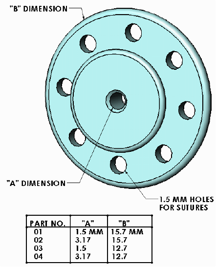
The stent used is machined from solid virgin Teflon® by McAllister Technical Services at West 280 Prairie Avenue, Coeur d'Alene, ID 83815, tel: 1-208-772-9527. The unit is 12.7 or 16 mm in diameter with eight pre-drilled holes for suture placement in the subcutaneous flange. The central stem is 1.5 mm deep and 4.5 mm in diameter with a 1.5 mm orifice. The outer rim is 8 mm in diameter, allowing the skin to tuck under the edge and thereby preventing the healing dermis from closing over the implant.
The surgery is performed under general anesthesia. Isoforane® induction and maintenance by mask and/or intubation is the agent and technique of choice. The surgical site is plucked of feathers, prepped and draped. The pre-autoclaved stent is prepared by placing four stainless steel wire lengths through the four pairs of pre-drilled holes in the flange. An incision is made through the skin just long enough for the stent to be slipped through and positioned under the skin with one set of holes straddling each end of the incision and one set of holes on each side of the incision. A twenty-two gauge hypodermic needle is used to retrieve the wire suture ends through the skin and the sutures are tied, both closing the incision and affixing the stent against the subcutaneous skin surface. The sutures are trimmed at their square knots and left in place. As healing occurs, the knots bury in the dermis.
The stents are inert and non-reactive. One post-surgical problem has been occasional plugging of the stent with mucous or congealed tissue fluid which is easily removed with a needle. This has occurred a few times post-operatively in some cases, but this problem eventually clears. Post-operative antibiotics have not been used and infected surgical sites have not occurred. To prevent post-surgical plugging in larger psittacines, it might be useful to use a larger stent with a 3 mm orifice and a 16 mm diameter external flange.
- Case 1: An 8-10 year old Mollucan cockatoo (Cacatua moluccensis) was presented in September 1990 for massive subcutaneous emphysema over the shoulders, thorax and neck. The onset occurred after the October 1989 San Francisco area earthquake. The bird had apparently been thrown around in its cage. Prior to presentation, the bird had been seen at another practice and the trapped air was drained with a hypodermic needle every thirty days over a six to eight month period. The bird was placed under Isoforane® anesthesia, the area on the dorsum of the neck just caudal to the skull was prepped and a stent implanted. No collar was placed on the bird post-operatively.
- Case 2: An orange-wing Amazon (Amazona amazonica) was presented with a subcutaneous air pocket involving the right cheek. The duration had been approximately four years. The bird persistently scratched the area which was denuded of feathers over the central portion. A Teflon® stent was implanted and because of chronic self-mutilation a 20 cm "buster collar" was placed around the neck. This stent plugged six times during the four months following placement of the stent, but by the ninth month post-operatively the subcutaneous air pocket was absent, the bird was comfortable and the area was totally refeathered.
Conclusion: Teflon® stents appear to be a clinically viable method of correcting subcutaneous emphysema in avian species.
Copyright© Association of Avian Veterinarians. Reprinted by permission.
Teflon® is a registered trademark of DuPont
*Formerly of Montclair Veterinary Clinic and Hospital
1961 Mountain Boulevard
Oakland, CA 94611, United States of America
Links to other Avian sites
- Up At Six Aviaries
- Kurt Thams Bird Cage Home Page
- Mark Bittner's page about the wild parrots of Telegraph Hill, in San Francisco